this post was submitted on 14 Jan 2025
814 points (100.0% liked)
pics
19927 readers
859 users here now
Rules:
1.. Please mark original photos with [OC] in the title if you're the photographer
2..Pictures containing a politician from any country or planet are prohibited, this is a community voted on rule.
3.. Image must be a photograph, no AI or digital art.
4.. No NSFW/Cosplay/Spam/Trolling images.
5.. Be civil. No racism or bigotry.
Photo of the Week Rule(s):
1.. On Fridays, the most upvoted original, marked [OC], photo posted between Friday and Thursday will be the next week's banner and featured photo.
2.. The weekly photos will be saved for an end of the year run off.
Instance-wide rules always apply. https://mastodon.world/about
founded 2 years ago
MODERATORS
you are viewing a single comment's thread
view the rest of the comments
view the rest of the comments


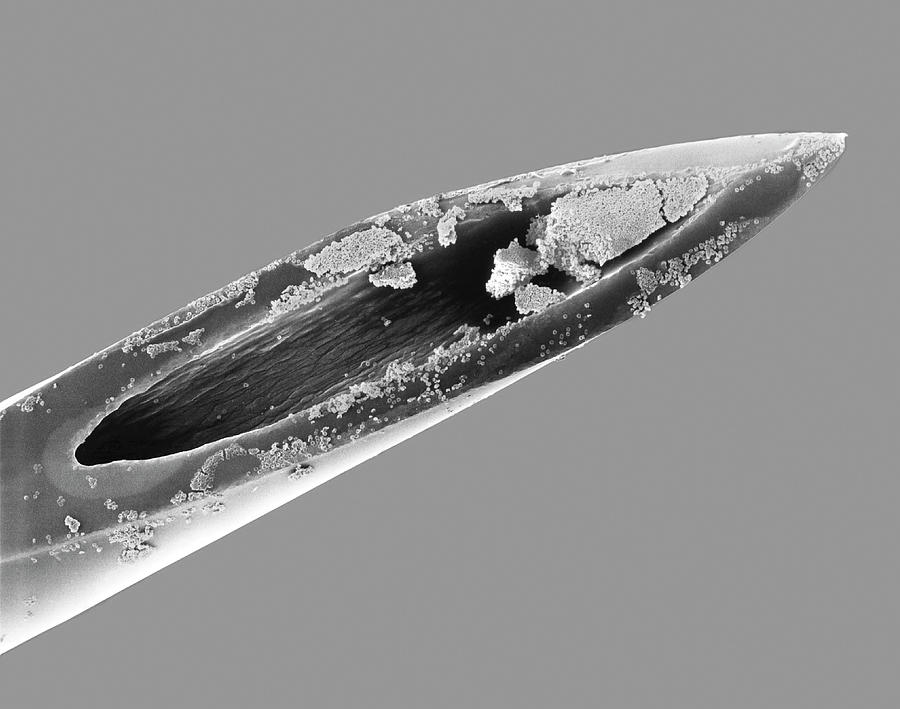
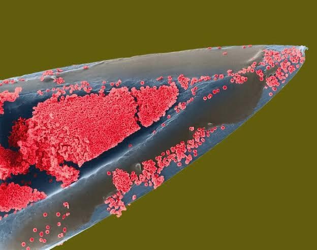
I actually thought optical microscopy worked just fine at this scale.
I know it's not the case for this photo, but if a 7 μm red bloodcell is reflecting red light (700nm, aka 0.7μm) under a bright white light, wouldn't the smallest discernable detail of the red blood cells be about a 10th of its width? Is that not roughly the detail we have in this image?
I'm making an assumption that the distance between discernable parts roughly parallels the wavelength's width. I could be wrong tho